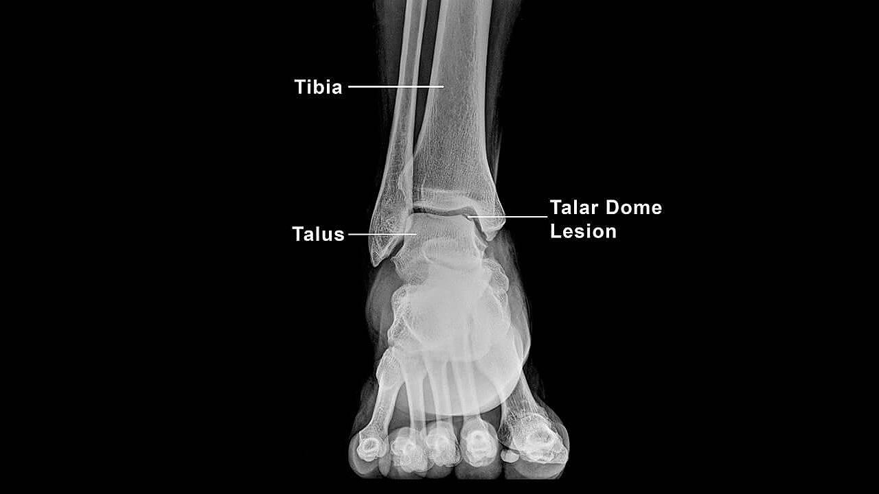
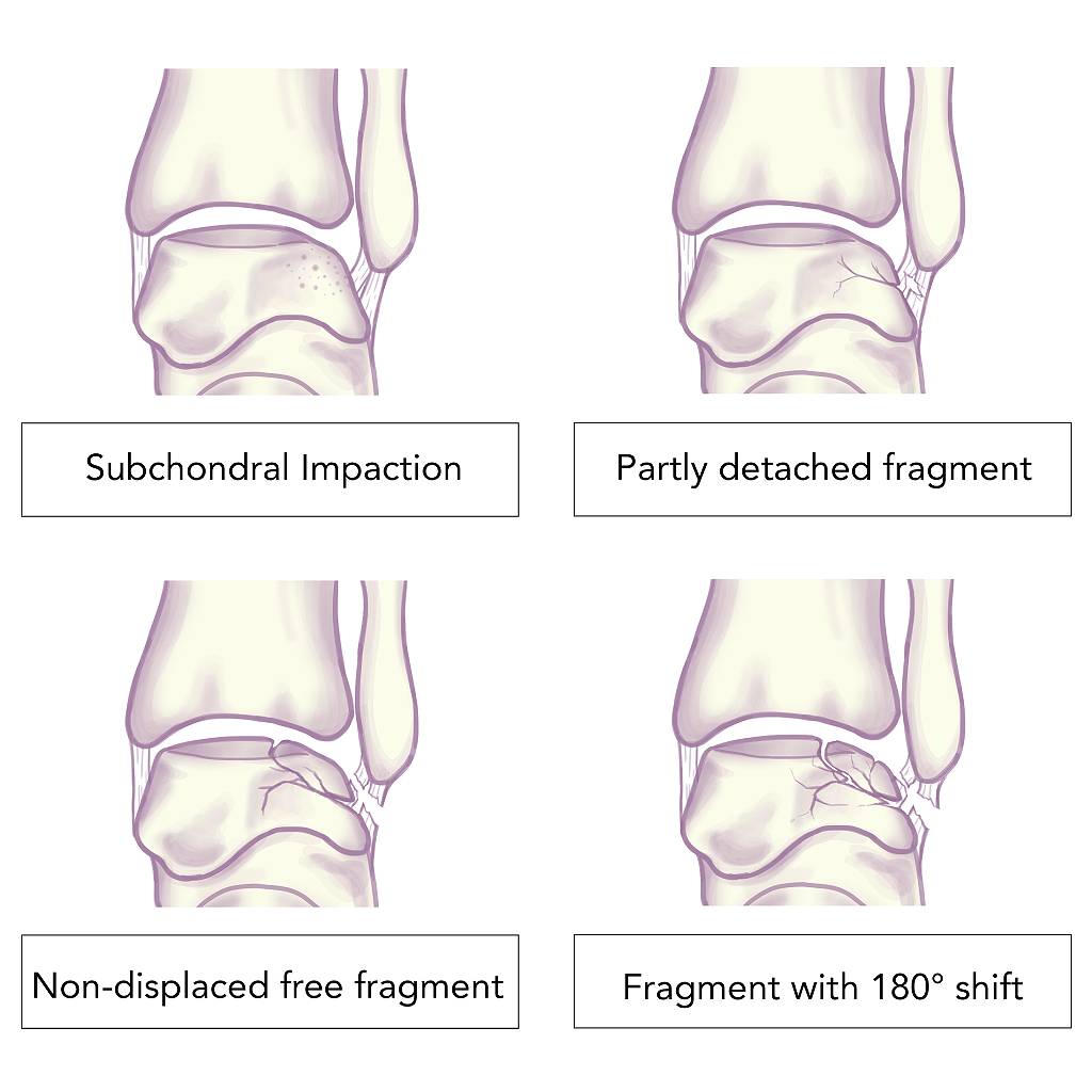
The talus bone, which is the part of the ankle that enables smooth movement, has a dome-shaped top covered in a layer of cartilage. An injury can cause damage to this area and the underlying subchondral bone, resulting in a talar dome injury typically referred to as an osteochondral lesion or defect. While injuries commonly present on the inside or medial aspect of the ankle, the outer ankle can be affected as well.
Causes of Talar Dome Injuries
The cause of a talar dome injury may be due to major traumatic events or repetitive stress injuries such as:
- Direct impact from heel strike
- Severe or recurrent ankle sprains
- Fractures
- Repetitive micro-trauma
Up to 70% of acute injuries like ankle sprains and fractures result in talar dome lesions1 If adequate care is not received, the condition may lead to improper healing, an increased risk of deterioration or softening of the cartilage, and further damage to the talus surface over time.
Continuous minor trauma over a prolonged period caused by unaddressed or undetected biomechanical issues can result in injury to the talar dome. These issues include hypermobility or a low-arch foot type (flatfoot), which can eventually lead to osteochondral lesions in both ankles.
Symptoms and Signs of Talar Dome Injury
Symptoms often develop gradually over time and may not be immediately noticeable, leading to misdiagnosis. These symptoms may present as:
- Chronic pain felt deep within the ankle
- Incidents of unprompted swelling, weakness, and locking of the ankle joint
- Formation of subchondral cysts
As the condition progresses, symptoms may worsen and restrict your lifestyle or impact your daily activities, especially if you are physically active. If left unresolved, this can commonly lead to osteoarthritis, changes in the ankle joint, recurrent and chronic pain linked to secondary nerve pain, and limited range of motion in the joint. Therefore, we recommend seeking proper solutions to prevent the onset of further debilitating symptoms.
Managing Talar Dome Injury
X-rays or an MRI of the ankle are performed to diagnose and determine the size, extent, and stability of the talar dome lesion or injury. The application of non-invasive podiatric intervention depends on several factors:
- Age
- Lesion size, extent, and stability
- Activity levels of patient
- Underlying biomechanical variances
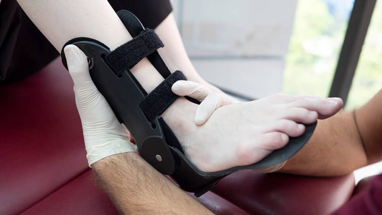
After this, patients may benefit from using offloading devices such as customised ankle foot orthotics (AFOs). AFOs can help to prevent the recurrence of previous injuries by stabilizing the ankle and preventing it from over-rotating. Your podiatrist will conduct follow-up appointments to ensure that the injury is healing and that the patient’s concerns have been addressed.
Surgery may be an option if non-invasive methods are ineffective in relieving pain or other related symptoms, or if the lesion is too severe. Do speak to your podiatrist to determine whether this is suitable for you.